Home
Products
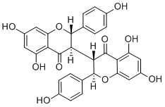
Isochamaejasmine


| Product Name | Isochamaejasmine |
| Price: | Inquiry |
| Catalog No.: | CN05101 |
| CAS No.: | 93859-63-3 |
| Molecular Formula: | C30H22O10 |
| Molecular Weight: | 542.5 g/mol |
| Purity: | >=98% |
| Type of Compound: | Flavonoids |
| Physical Desc.: | Powder |
| Source: | The roots of Stellera chamaejasme Linn. |
| Solvent: | Chloroform, Dichloromethane, Ethyl Acetate, DMSO, Acetone, etc. |
| SMILES: | Oc1ccc(cc1)C1Oc2cc(O)cc(c2C(=O)C1C1C(Oc2c(C1=O)c(O)cc(c2)O)c1ccc(cc1)O)O |
| Contact us | |
|---|---|
| First Name: | |
| Last Name: | |
| E-mail: | |
| Question: | |
| Description | Isochamaejasmin is a biflavonoid with anti-cancer, antiplasmodial and insecticidal activities. Isochamaejasmin displays a potent NF-κB (NF-κB) activation activity. Isochamaejasmin could cause DNA damage and induce Apoptosis via the mitochondrial pathway in AW1 cells[1][2]. Isochamaejasmin also has a moderate antiplasmodial activity (IC50 of 7.3 μM for P. falciparum) and relatively low cytotoxicity (CC50 of 29.0 μM)[3]. |
| In Vitro | Isochamaejasmin (6.25-100μM; 24-72 h) 通过时间和剂量依赖性方式显示出对 AW1 细胞的潜在毒性。Isochamaejasmin (1000 mg/L, 500 mg/L, 250 mg/L, 125 mg/L, 62.5 mg/L; 24 h, 48 h, 72 h, and 96 h) 对 H. zea 幼虫具有随时间的潜在毒性和剂量依赖性行为[1]。 Isochamaejasmin (40-80μM; 24 h) 导致 DNA 损伤并增加 AW1 细胞中 γH2AX 和 OGG1 的水平。使细胞周期停滞在G2/M期[1]。 Isochamaejasmin (20-80μM; 24 h) 剂量依赖性诱导 AW1 细胞凋亡[1] 。 Isochamaejasmin (20-80μM; 24 h) 显示 MMP 下降,Bax/Bcl-2 表达上调,导致细胞色素 c 释放到胞质溶胶中,caspase-3/9 激活,PARP 裂解 [1] 。 Isochamaejasmin 显示 AW1 细胞中活性氧 (ROS) 水平的剂量依赖性升高、脂质过氧化产物的积累和抗氧化酶的失活[1]。 Isochamaejasmin 在转染的 HeLa 细胞中诱导 NF-κB 定向报告基因的表达,EC50 为 3.23 μM。Isochamaejasmin 刺激的 NF-κB 报告基因活性伴随着 NF-κB 蛋白的核转位,并被 IκBα 的显性失活结构阻断。Isochamaejasmin 还诱导丝裂原活化蛋白激酶 ERK1/2 和 p38 以及 PKCδ的时间依赖性磷酸化[2] 。 Cell Viability Assay[1] Cell Line: Larvae and neuronal cells (AW1) Concentration: 6.25 μM, 12.5 μM, 25 μM, 50 μM and 100 μM Incubation Time: 24 h, 48 h and 72 h Result: Had potential toxicity against H. zea both in vivo and in vitro via time- and dose-dependent manners. Cell Cycle Analysis[1] Cell Line: Neuronal cells (AW1) Concentration: 40 μM and 80 μM Incubation Time: 24 h Result: The cell cycle was arrested at the G2/M phase. Apoptosis Analysis[1] Cell Line: Neuronal cells (AW1) Concentration: 20 μM, 40 μM, and 80 μM Incubation Time: 24 h Result: Induced apoptosis via the mitochondrial pathway in AW1 cells. Western Blot Analysis[1] Cell Line: Neuronal cells (AW1) Concentration: 20 μM, 40 μM, and 80 μM Incubation Time: 24 h Result: Showed upregulation of Bax/Bcl-2 expression resulting in the release of cytochrome c into the cytosol, activation of caspase-3/9, and cleavage of PARP. |
| Density | 1.6±0.1 g/cm3 |
| Boiling Point | 932.6±65.0 °C at 760 mmHg |
| Flash Point | 314.2±27.8 °C |
| Exact Mass | 542.121277 |
| PSA | 173.98000 |
| LogP | 6.61 |
| Vapour Pressure | 0.0±0.3 mmHg at 25°C |