Home
Products
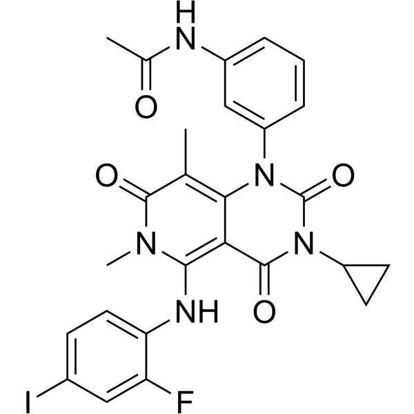
Trametinib (GSK1120212)


| Product Name | Trametinib (GSK1120212) |
| Price: | Inquiry |
| Catalog No.: | CN00514 |
| CAS No.: | 871700-17-3 |
| Molecular Formula: | C26H23FIN5O4 |
| Molecular Weight: | 615.39 g/mol |
| Purity: | >=98% |
| Type of Compound: | Alkaloids |
| Physical Desc.: | Powder |
| Source: | |
| Solvent: | Chloroform, Dichloromethane, Ethyl Acetate, DMSO, Acetone, etc. |
| SMILES: | CC(=O)Nc1cccc(c1)n1c(=O)n(C2CC2)c(=O)c2c1c(C)c(=O)n(c2Nc1ccc(cc1F)I)C |
| Contact us | |
|---|---|
| First Name: | |
| Last Name: | |
| E-mail: | |
| Question: | |
| Description | Trametinib is a potent MEK inhibitor that inhibits MEK1 and MEK2 with IC50s of about 2 nM. Due to the poor solubility of Trametinib, Trametinib DMSO solvate (Cat. No.: HY-10999A) is recommeded. |
| Target | MEK1:2 nM (IC50) MEK2:2 nM (IC50) |
| In Vitro | Trametinib (0.1-100 nM) blocks tumor necrosis factor-α and interleukin-6 production from peripheral blood mononuclear cells (PBMCs). Trametinib (JTP-74057) inhibits the growth of 9 out of 10 human colorectal cancer cell lines, and they shows cell-cycle arrest at the G1 phase after drug tratment[1]. The combination of GSK2118436 and Trametinib (GSK1120212) effectively inhibits cell growth, decreases ERK phosphorylation, decreases cyclin D1 protein, and increases p27(kip1) protein in the resistant clones[2]. |
| In Vivo | Adjuvant-induced arthritis (AIA) and type II collageninduced arthritis (CIA) development are suppressed almost completely by 0.1 mg/kg of Trametinib (JTP-74057) or 10 mg/kg of Leflunomide[1]. Trametinib (0.3 mg/kg, 1 mg/kg, p.o.) is effective in inhibiting the HT-29 xenograft growth in a nude mouse xenograft model[2]. |
| Cell Assay | Cells are treated with various concentrations of Trametinib (JTP-74057) in 100 mm dishes for 3 or 4 days. Both floating and adherent cells are collected and fixed with 70% ethanol. After washing with PBS, the cells are suspended in 100 µL/mL RNase and 25 µL/mL Propidium iodide (PI) and incubated at 37°C for 30 min in the dark. The DNA content of each single cell is determined using the flow cytometer Cytomics FC500 or Guava EasyCyte plus[2]. |
| Animal Admin | Mice[2] Female BALB/c-nu/nu mice are used. On day 0, HT-29 cells or COLO205 cells suspended in ice-cold HBSS (-) are inoculated subcutaneously into the right flank of the mice at 5×10⁶ cells/100 µL/site or 1×10⁶ cells/100 µL/site, respectively. The acetic acid-solvated form of Trametinib (JTP-74057, 0.3 mg/kg, 1 mg/kg) is dissolved in 10% Cremophor EL-10% PEG400 and is administered orally once daily for 14 days from the day when the mean tumor volume reached 100 mm3. The tumor length [L(mm)] and width [W(mm)] are measured using a microgauge twice a week after commencement of dosing, and the tumor volume is calculated using the following formula: tumor volume (mm3)=L×W×W/2. |
| Density | 1.7±0.1 g/cm3 |
| Exact Mass | 615.077881 |
| PSA | 110.62000 |
| LogP | 2.68 |
| Storage condition | 2-8°C |