Home
Products
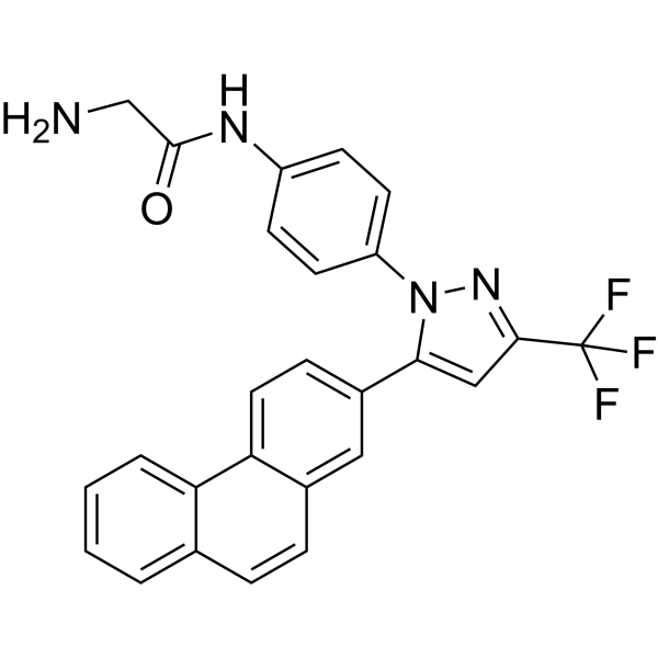
OSU-03012 (AR-12)


| Product Name | OSU-03012 (AR-12) |
| Price: | Inquiry |
| Catalog No.: | CN00367 |
| CAS No.: | 742112-33-0 |
| Molecular Formula: | C26H19F3N4O |
| Molecular Weight: | 460.45 g/mol |
| Purity: | >=98% |
| Type of Compound: | Alkaloids |
| Physical Desc.: | Powder |
| Source: | |
| Solvent: | Chloroform, Dichloromethane, Ethyl Acetate, DMSO, Acetone, etc. |
| SMILES: | NCC(=O)Nc1ccc(cc1)n1nc(cc1c1ccc2c(c1)ccc1c2cccc1)C(F)(F)F |
| Contact us | |
|---|---|
| First Name: | |
| Last Name: | |
| E-mail: | |
| Question: | |
| Description | OSU-03012 is a PDK-1 inhibitor with an IC50 of 5 μM. |
| Target | IC50: 5 μM (PDK-1)[1] |
| In Vitro | OSU-03012 inhibits PC-3 cells viability with IC50 values of 5 μM. The effects of OSU-03012 on PC-3 cell proliferation in 10% FBS-supplemented medium are also examined. OSU-03012 induces apoptotic death in PC-3 cells in 1% FBS-containing medium in a dose-dependent manner, as demonstrated by DNA fragmentation and PARP cleavage. OSU-03012 is effective in suppressing PC-3 cell proliferation at sub-μM, consistent with that noted in 1% serum[1]. |
| In Vivo | All of the SCID/Rag2 mice develop two MDA-MB-435/LCC6/Her-2 tumors and are assigned to either the vehicle control or OSU-03012 (200 mg/kg) treatment group, which is given orally for 3 days. OSU-03012 remarkably decreases EGFR protein expression in the tumors by ~48% compared with expression levels found in the tumors taken from mice that receive the vehicle control. OSU-03012 also prevents Y-box binding protein-1 (YB-1) from binding to the EGFR promoter at the 1b and 2a sites[2]. |
| Cell Assay | PC-3 (p53-/-) human androgen-nonresponsive prostate cancer cells are cultured in RPMI 1640 supplemented with 10% fetal bovine serum (FBS) at 37°C in a humidified incubator containing 5% CO2. The effect of Celecoxib and its derivatives (e.g., OSU-03012) (2.5 μM, 5 μM, 7.5 μM and 10 μM) on PC-3 cell viability is assessed by using the MTT assay in six replicates. Cells are grown in 10% FBS- supplemented RPMI 1640 in 96-well, flat-bottomed plates for 24 h, and are exposed to various concentrations of Celecoxib derivatives (e.g., OSU-03012) dissolved in DMSO (final concentration ≤0.1%) in 1% serum-containing RPMI 1640 for different time intervals. Controls receive DMSO vehicle at a concentration equal to that in drug-treated cells. The medium is removed, replaced by 200 μL of 0.5 mg/mL of MTT in 10% FBS-containing RPMI 1640, and cells are incubated in the CO2 incubator at 37°C for 2 h. Supernatants are removed from the wells, and the reduced MTT dye is solubilized in 200 μL/well DMSO. Absorbance at 570 nm is determined on a plate reader[1]. |
| Animal Admin | Mice[2] SCID/Rag2m mice (6-8 weeks old, female) are subcutaneously injected with 1×10⁷ MDA-MB-435/LCC6 cells stably transfected with HER-2/neu. Each mouse is inoculated with the cells on the right and left sides of the lower back. A total of eight mice are injected, each harboring two tumors. After 6 weeks, the mice are randomly assigned into groups (vehicle, 0.5% methyl cellulose/0.1% Tween 80, or OSU-03012 at 200 mg/kg/day). Mice are dosed daily for 3 days with either the vehicle or OSU-03012 by oral gavage. On the fourth day, the study is terminated, mice are sacrificed, and the tumors are collected for chromatin immunoprecipitation (ChIP) and protein isolations. |
| Density | 1.4±0.1 g/cm3 |
| Boiling Point | 683.0±55.0 °C at 760 mmHg |
| Flash Point | 366.9±31.5 °C |
| Exact Mass | 460.151093 |
| PSA | 72.94000 |
| LogP | 5.38 |
| Vapour Pressure | 0.0±2.1 mmHg at 25°C |
| Storage condition | -20°C |