Home
Products
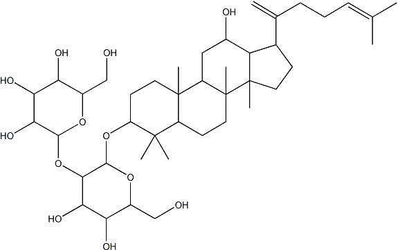
Ginsenoside Rk1


| Product Name | Ginsenoside Rk1 |
| Price: | $161 / 10mg |
| Catalog No.: | CN07614 |
| CAS No.: | 494753-69-4 |
| Molecular Formula: | C42H70O12 |
| Molecular Weight: | 767.0 g/mol |
| Purity: | >=98% |
| Type of Compound: | Triterpenoids |
| Physical Desc.: | Powder |
| Source: | The roots of Panax ginseng C. A. Mey. |
| Solvent: | DMSO, Pyridine, Methanol, Ethanol, etc. |
| SMILES: | OC[C@H]1O[C@@H](O[C@H]2CC[C@]3([C@H](C2(C)C)CC[C@@]2([C@@H]3C[C@@H](O)[C@H]3[C@@]2(C)CC[C@@H]3C(=C)CCC=C(C)C)C)C)[C@@H]([C@H]([C@@H]1O)O)O[C@@H]1O[C@H](CO)[C@H]([C@@H]([C@H]1O)O)O |
| Contact us | |
|---|---|
| First Name: | |
| Last Name: | |
| E-mail: | |
| Question: | |
| Description | Ginsenoside Rk1 is a unique component created by processing the ginseng plant (mainly Sung Ginseng, SG) at high temperatures[1].Ginsenoside Rk1 has anti-inflammatory effect, suppresses the activation of Jak2/Stat3 signaling pathway and NF-κB[2].Ginsenoside Rk1 has anti-tumor effect, antiplatelet aggregation activities, anti-insulin resistance, nephroprotective effect, antimicrobial effect, cognitive function enhancement, lipid accumulation reduction and prevents osteoporosis[1].Ginsenoside Rk1 induces cell apoptosis by triggering intracellular reactive oxygen species (ROS) generation and blocking PI3K/Akt pathway[3]. |
| In Vitro | Ginsenoside Rk1 (0-40 μM; 6 hours) inhibits MCP-1 and TNF-α mRNA induced by lipopolysaccharide (LPS), expression of IL-1β is inhibited at 40 μM[2]. Ginsenoside Rk1 (0-40 μM; 24 hours) inhibits phosphorylation of JAK2 and STAT3 (Tyr705 and Ser727) in LPS-induced RAW264.7 cells in a dose-dependent manner[2]. Ginsenoside Rk1 (0-160 μM; 48 hours) results in cell viability significant decrease 75.52 ± 2.51% (40 μM), 52.72 ± 2.54% (80 μM), 17.41 ± 2.94% (120 μM)and 12.63 ± 3.24% (160 μM) compared with control[3]. Ginsenoside Rk1 (0-120 μM; 24 hours) increases G0/G1 phase proportion accompanied with S and G2/M phase proportion decrease in MDA-MB-231 cells[3]. Ginsenoside Rk1 (0-120 μM; 24 hours) promotes the percentage of apoptotic cells in a dose-dependent manner, exhibits to reduction of cell number with nucleus fragmentation, condensation and apoptotic body formation[3]. RT-PCR[2] Cell Line: RAW264.7 cells Concentration: 10 μM, 20 μM, 40 μM Incubation Time: 6 hours Result: Inhibited JAK2-dependent STAT3 activation in LPS-activated RAW264.7 cells. Western Blot Analysis[2] Cell Line: RAW264.7 cells Concentration: 10 μM, 20 μM, 40 μM Incubation Time: 6 hours Result: Inhibited JAK2-dependent STAT3 activation in LPS-activated RAW264.7 cells Cell Viability Assay[3] Cell Line: MDA-MB-231 cells Concentration: 0 μM, 40 μM, 80 μM, 120 μM Incubation Time: 48 hours Result: Inhibited MDA-MB-231 cells proliferation in a dose- and time-dependent manner. Cell Cycle Analysis[3] Cell Line: MDA-MB-231 cells Concentration: 0 μM, 40 μM, 80 μM, 120 μM Incubation Time: 24 hours Result: Induced G0/G1 phase arrest. Apoptosis Analysis[3] Cell Line: MDA-MB-231 cells Concentration: 0 μM, 40 μM, 80 μM, 120 μM Incubation Time: 24 hours Result: Induced apoptosis in MDA-MB-231 cells. |
| Density | 1.3±0.1 g/cm3 |
| Boiling Point | 854.5±65.0 °C at 760 mmHg |
| Flash Point | 470.6±34.3 °C |
| Exact Mass | 766.486755 |
| PSA | 198.76000 |
| LogP | 6.70 |
| Vapour Pressure | 0.0±0.6 mmHg at 25°C |
| Storage condition | -20℃ |