Home
Products
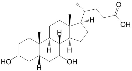
Chenodeoxycholic acid


| Product Name | Chenodeoxycholic acid |
| Price: | $15 / 20mg |
| Catalog No.: | CN02458 |
| CAS No.: | 474-25-9 |
| Molecular Formula: | C24H40O4 |
| Molecular Weight: | 392.57 g/mol |
| Purity: | >=98% |
| Type of Compound: | Steroids |
| Physical Desc.: | Cryst. |
| Source: | From the bile. |
| Solvent: | Chloroform, Dichloromethane, Ethyl Acetate, DMSO, Acetone, etc. |
| SMILES: | O[C@@H]1CC[C@]2([C@@H](C1)C[C@H]([C@@H]1[C@@H]2CC[C@]2([C@H]1CC[C@@H]2[C@@H](CCC(=O)O)C)C)O)C |
| Contact us | |
|---|---|
| First Name: | |
| Last Name: | |
| E-mail: | |
| Question: | |
| Description | Chenodeoxycholic Acid is a hydrophobic primary bile acid that activates nuclear receptors (FXR) involved in cholesterol metabolism. |
| Target | Human Endogenous Metabolite |
| In Vitro | Chenodeoxycholic acid (CDCA) and Deoxycholic acid (DCA) both inhibit 11 beta HSD2 with IC50 values of 22 mM and 38 mM, respectively and causes cortisol-dependent nuclear translocation and increases transcriptionalactivity of mineralocorticoid receptor (MR)[1]. Chenodeoxycholic acid is able to stimulate Ishikawa cell growth by inducing a significant increase in Cyclin D1 protein and mRNA expression through the activation of the membrane G protein-coupled receptor (TGR5)-dependent pathway[2]. Chenodeoxycholic acid (CDCA) induces LDL receptor mRNA levels approximately 4 fold and mRNA levels for HMG-CoA reductase and HMG-CoA synthase two fold in a cultured human hepatoblastoma cell line, Hep G2[3]. Chenodeoxycholic acid-induced Isc is inhibited (≥67%) by Bumetanide, BaCl2, and the cystic fibrosis transmembrane conductance regulator (CFTR) inhibitor CFTRinh-172. Chenodeoxycholic acid-stimulated Isc is decreased 43% by the adenylate cyclase inhibitor MDL12330A and Chenodeoxycholic acid increases intracellular cAMP concentration[4]. Chenodeoxycholic acid treatment activates C/EBPβ, as shown by increases in its phosphorylation, nuclear accumulation, and expression in HepG2 cells. Chenodeoxycholic acid enhances luciferase gene transcription from the construct containing -1.65-kb GSTA2 promoter, which contains C/EBP response element (pGL-1651). Chenodeoxycholic acid treatment activates AMP-activated protein kinase (AMPK), which leads to extracellular signal-regulated kinase 1/2 (ERK1/2) activation, as evidenced by the results of experiments using a dominant-negative mutant of AMPKα and chemical inhibitor[5]. |
| Cell Assay | The cell viability is analyzed by incubating transfected HEK-293 cells and CHO cells for 1 h with the corresponding concentration of bile acid and staining with trypan blue. The toxicity of bile acids is analyzed using the tetrazolium salt MTT (3-(4,5-dimethylthiazol-2-yl)-2,5-diphenyltetrazolium bromide) according to the cell proliferation kit I. No significant differences between control and bile acid-treated cells are obtained in both tests. |
| Density | 1.1±0.1 g/cm3 |
| Boiling Point | 547.1±25.0 °C at 760 mmHg |
| Flash Point | 298.8±19.7 °C |
| Exact Mass | 392.292664 |
| PSA | 77.76000 |
| LogP | 4.66 |
| Vapour Pressure | 0.0±3.3 mmHg at 25°C |