Home
Products
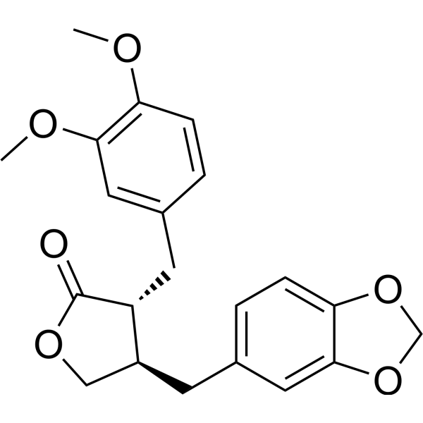
Bursehernin


| Product Name | Bursehernin |
| Price: | Inquiry |
| Catalog No.: | CN02884 |
| CAS No.: | 40456-51-7 |
| Molecular Formula: | C21H22O6 |
| Molecular Weight: | 370.4 g/mol |
| Purity: | >=98% |
| Type of Compound: | Lignans |
| Physical Desc.: | Powder |
| Source: | The herbs of Macaranga tanarius |
| Solvent: | Chloroform, Dichloromethane, Ethyl Acetate, DMSO, Acetone, etc. |
| SMILES: |
| Contact us | |
|---|---|
| First Name: | |
| Last Name: | |
| E-mail: | |
| Question: | |
| Description | Bursehernin (Methylpluviatolide) is an antitumor agent. Bursehernin induces Apoptosis and cell cycle arrest at G2/M phase. Bursehernin shows anti-proliferative activity[1][2]. |
| In Vitro | Bursehernin (4.3 µM for MCF-7 cells, 3.7 µM for KKU-M213 cells; 4, 48, 72 h) 以时间依赖的方式诱导细胞凋亡和细胞周期停滞在G2/M期 [1]。 Cell Proliferation Assay [1] Cell Line: MCF-7, MDA-MB-468, MDA-MB-231, HT-29, KKU-M213, KKU-K100, KKU-M055, L-929, MMNK-1 cells Concentration: 0-100 µM Incubation Time: 72 h Result: Showed anti-proliferative activity with IC50s of 11.96, 8.24, 14.26, 47.53, 3.70, 12.38, 17.38, 26.36, 7.45 µM for MCF-7, MDA-MB-468, MDA-MB-231, HT-29, KKU-M213, KKU-K100, KKU-M055, L-929, MMNK-1 cells, respectively. Cell Cycle Analysis [1] Cell Line: MCF-7, KKU-M213 cells Concentration: 4.3 µM for MCF-7 cells, 3.7 µM for KKU-M213 cells Incubation Time: 24, 48, 72 h Result: Induced cell cycle arrest at G2/M phase. Western Blot Analysis [1] Cell Line: MCF-7, KKU-M213 cells Concentration: 4.3 µM for MCF-7 cells, 3.7 µM for KKU-M213 cells Incubation Time: 24, 48, 72 h Result: Decreased the expression of topoisomerase II, STAT 3, cyclin D1, and p21. Apoptosis Analysis [1] Cell Line: MCF-7 cells Concentration: 0, 2.15, 4.30, 8.60 µM Incubation Time: 24, 48, 72, 96 h Result: Induced apoptosis in a time- and dose-dependent manner. |
| Density | 1.268g/cm3 |
| Boiling Point | 543.575°C at 760 mmHg |
| Flash Point | 238.539 °C |
| Exact Mass | 370.14200 |
| PSA | 63.22000 |
| LogP | 3.00690 |