Home
Products
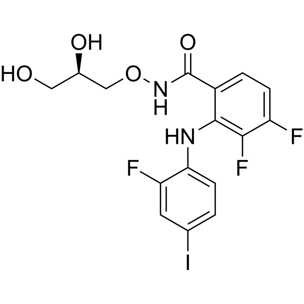
PD0325901(Mirdametinib)


| Product Name | PD0325901(Mirdametinib) |
| Price: | Inquiry |
| Catalog No.: | CN00533 |
| CAS No.: | 391210-10-9 |
| Molecular Formula: | C16H14F3IN2O4 |
| Molecular Weight: | 482.19 g/mol |
| Purity: | >=98% |
| Type of Compound: | Alkaloids |
| Physical Desc.: | Powder |
| Source: | |
| Solvent: | Chloroform, Dichloromethane, Ethyl Acetate, DMSO, Acetone, etc. |
| SMILES: | OC[C@H](CONC(=O)c1ccc(c(c1Nc1ccc(cc1F)I)F)F)O |
| Contact us | |
|---|---|
| First Name: | |
| Last Name: | |
| E-mail: | |
| Question: | |
| Description | PD0325901 is a selective and cell permeable MEK inhibitor with an IC50 of 0.33 nM. |
| Target | MEK1:0.33 nM (IC50) Autophagy |
| In Vitro | PF0325901 shows higher permeability, and should be able to achieve higher systemic exposures than CI-1040[1]. PD0325901 is exquisitely specific and highly potent against purified MEK, revealing a Kiapp of 1 nM against activated MEK1 and MEK2. PD0325901 is roughly 500-fold more potent than CI-1040 with respect to its cellular effects on phosphorylation of ERK1 and ERK2, displaying subnanomolar activity. PD0325901 prevents the growth of melanoma cell lines. PD0325901 inhibits the growth of TPC-1 cells and K2 cells with GC50 of 11 nM and 6.3 nM, respectively. PD0325901 significantly prevents the the growth of PTC cells harboring a BRAF mutation at very low concentration (10 nM) and only moderately increases the growth of the PTC cells carrying the RET/PTC1 rearrangement at the same concentration. PD0325901 effectively inhibits the phosphorylation of ERK1/2 in multiple PTC cell lines[2]. |
| In Vivo | PD0325901 (25 mg/kg, p.o.) inhibits phosphorylation of ERK by more than 50% at 24 hours post-dosing. The dose required to produce a 70% incidence of complete tumor responses (C26 model) is 25 mg/kg/day versus 900 mg/kg/day for PD0325901 and CI-1040, respectively. Anticancer activity of PD 0325901 has been demonstrated for a broad spectrum of human tumor xenografts. PD0325901 (20-25 mg/kg/day, p.o.) treatment in mice, shows no tumor growth inoculated with PTC cells bearing a BRAF mutation. For PTC with the RET/PTC1 rearrangement, the average tumor volume of the orthotopic tumor is decreased by 58% as compared with controls. PTC cells carrying a BRAF mutation are more sensitive to PD0325901 than are PTC cells carrying the RET/PTC1 rearrangement[2]. |
| Cell Assay | PTC cells (1×10⁴) are seeded in 24-well plates with 1 mL of medium for 4 days in a 37°C incubator. MEK inhibitor PD0325901 at varying concentrations is added to the cells in triplicate on day 0. MTT dissolved in 0.8% NaCl solution at 5 mg/mL is added to each well (0.2 mL) on day 2 to test GC50 or every day for cell growth curves. The cells are incubated at 37°C for 3 hours with MTT. The liquid is then aspirated from the wells and discarded. Stained cells are dissolved in 0.5 mL of DMSO and their absorption at 570 nm is measured using a Synergy HT multidetection microplate reader. For GC50, cell growth is calculated as 100×(T−T0)/(C−T0), where T is the optical density of the wells treated with inhibitors after a 48-hour period, T0 is the optical density at time zero, and C is the control optical density with DMSO only. |
| Animal Admin | Mice (10-14 per group) are anesthetized s.c. with a cocktail (100 μL/10 g body weight of 10 mg/mL keta-mine and 1 mg/mL xylazine). K2 and TPC-1 cells stably infected with a retrovirus expressing luciferase (5×10⁵ cells in 5 μL RPMI1640 medium) are inoculated into the thyroid gland, and the mice are monitored weekly for tumor growth by Xenogen using Living Image 3.0 software. One week after inoculation, PD0325901 is dissolved in 80 mM citric buffer (pH 7) by sonication and given to mice daily by oral gavage (20-25 mg/kg) for 3 weeks (5 consecutive days/week). Mice are sacrificed only due to tumor burden or loss of 20% of body weight. Tumor sizes are measured with calipers and tumor volume (V) is calculated by the formula (V=length×width×depth). Control mice are given 80 mM citric buffer (pH 7) alone. All in vivo experiments are done at least twice. |
| Density | 1.8±0.1 g/cm3 |
| Exact Mass | 481.995026 |
| PSA | 90.82000 |
| LogP | 6.16 |
| Storage condition | Store at -20°C |