Home
Products
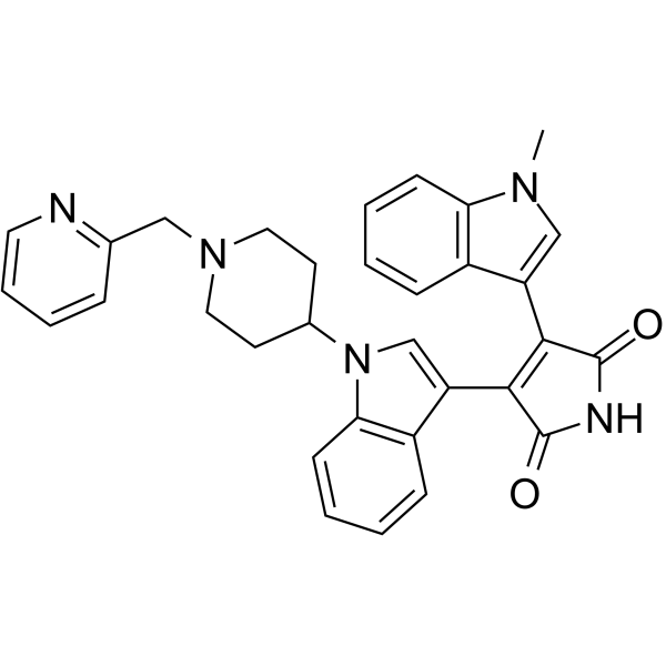
Enzastaurin (LY317615)


| Product Name | Enzastaurin (LY317615) |
| Price: | Inquiry |
| Catalog No.: | CN00441 |
| CAS No.: | 170364-57-5 |
| Molecular Formula: | C32H29N5O2 |
| Molecular Weight: | 515.61 g/mol |
| Purity: | >=98% |
| Type of Compound: | Alkaloids |
| Physical Desc.: | Powder |
| Source: | |
| Solvent: | Chloroform, Dichloromethane, Ethyl Acetate, DMSO, Acetone, etc. |
| SMILES: | O=C1NC(=O)C(=C1c1cn(c2c1cccc2)C1CCN(CC1)Cc1ccccn1)c1cn(c2c1cccc2)C |
| Contact us | |
|---|---|
| First Name: | |
| Last Name: | |
| E-mail: | |
| Question: | |
| Description | Enzastaurin is a potent and selective PKCβ inhibitor with an IC50 of 6 nM, showing 6- to 20-fold selectivity over PKCα, PKCγ and PKCε. |
| Target | PKCβ:6 nM (IC50) PKCα:39 nM (IC50) PKCγ:83 nM (IC50) PKCε:110 nM (IC50) |
| In Vitro | Enzastaurin increases apoptosis in malignant lymphocytes of CTCL. When combined with GSK3 inhibitors, enzastaurin demonstrates an enhancement of cytotoxicity levels. Treatment with a combination of enzastaurin and the GSK3 inhibitor AR-A014418 leads to increased levels of β-catenin total protein and β-catenin-mediated transcription. Blocking of β-catenin-mediated transcription or small hairpin RNA (shRNA) knockdown of β-catenin induces the same cytotoxic effects as that of enzastaurin plus AR-A014418. Additionally, treatment with enzastaurin and AR-A014418 decreases the mRNA levels and surface expression of CD44[1].Enzastaurin application results in a marked dose-dependent inhibition of growth in all MM cell lines investigated, including MM.1S, MM.1R, RPMI 8226 (RPMI), RPMI-Dox40 (Dox40), NCI-H929, KMS-11, OPM-2, and U266, with IC50 from 0.6-1.6 μM. Enzastaurin direct impacts human tumor cells, inducing apoptosis and suppressing proliferation in cultured tumor cells. Enzastaurin also suppresses the phosphorylation of GSK3βser9, ribosomal protein S6S240/244, and AKTThr308 while having no direct effect on VEGFR phosphorylation[3]. |
| In Vivo | Treatment of xenografts with Enzastaurin and radiation produces greater reductions in density of microvessels than either treatment alone. The decrease in microvessel density corresponds to delayed tumor growth[2]. |
| Cell Assay | Induction of apoptosis by enzastaurin is measured by nucleosomal fragmentation and terminal deoxynucleotidyl transferase-mediated nick-end labeling (TUNEL) and staining in HCT116 and U87MG cell lines. Briefly, 5×10³ cells are plated per well in 96-well plates (1% FBS-supplemented media conditions), incubated with or without Enzastaurin for 48 to 72 hours. The absorbance values are normalized to those from control-treated cells to derive a nucleosomal enrichment factor at all concentrations as per the manufacturer's protocol. The concentrations studied ranges from 0.1 to 10 μM. In situ TUNEL staining is assayed with the In situ Cell Death Detection, Fluorescein kit. Cells (7.5×10⁴) are plated per well in 6-well plates and incubated 72 hours in 1% FBS-supplemented media Enzastaurin. Fluorescein-labeled DNA strand breaks are detected with the BD epics flow cytometer. Ten thousand, single-cell, FITC-staining events are collected for each test. |
| Density | 1.34 |
| Boiling Point | 767.2±60.0 °C at 760 mmHg |
| Flash Point | 417.8±32.9 °C |
| Exact Mass | 515.232117 |
| PSA | 72.16000 |
| LogP | 4.43 |
| Vapour Pressure | 0.0±2.6 mmHg at 25°C |
| Storage condition | -20°C |