Home
Products
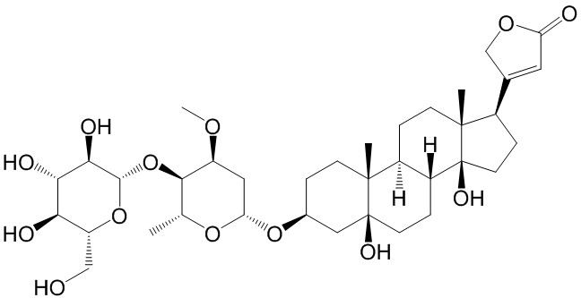
Periplocin


| Product Name | Periplocin |
| Price: | $62 / 20mg |
| Catalog No.: | CN02451 |
| CAS No.: | 13137-64-9 |
| Molecular Formula: | C36H56O13 |
| Molecular Weight: | 696.82 g/mol |
| Purity: | >=98% |
| Type of Compound: | Steroids |
| Physical Desc.: | Powder |
| Source: | The root barks of Periploca sepium Bunge. |
| Solvent: | DMSO, Pyridine, Methanol, Ethanol, etc. |
| SMILES: | CO[C@H]1C[C@H](O[C@H]2CC[C@]3([C@](C2)(O)CC[C@@H]2[C@@H]3CC[C@]3([C@]2(O)CC[C@@H]3C2=CC(=O)OC2)C)C)O[C@@H]([C@H]1O[C@@H]1O[C@H](CO)[C@H]([C@@H]([C@H]1O)O)O)C |
| Contact us | |
|---|---|
| First Name: | |
| Last Name: | |
| E-mail: | |
| Question: | |
| Description | Periplocin is a cardiotonic steroid isolated from Periploca forrestii. Periplocin promotes tumor cell apoptosis and inhibits tumor growth. Periplocin has the potential to facilitate wound healing through the activation of Src/ERK and PI3K/Akt pathways mediated by Na/K-ATPase[1][2]. |
| Target | Apoptosis[2] |
| In Vitro | Periplocin (5-20 μM; 48 hours; L929 cells) treatment shows increased proliferation up to 131% at 20 μM[1]. Periplocin (5-20 μM; 30-120 minutes; L929 cells) increases phosphorylation of Src, ERK, PI3K and Akt at active sites in a dosedependent and time-dependent manner.Periplocin (5-20 μM; 48 hours) significantly promotes migration of fibroblast cell[1]. Periplocin (5-20 μM; 48 hours) increases collagen production in L929 fibroblast[1]. Periplocin induces Na/KATPase mediates the activation of Src/ERK and PI3K/Akt pathways[1]. Cell Viability Assay[1] Cell Line: L929 cells Concentration: 5 μM, 10 μM, 20 μM Incubation Time: 48 hours Result: Showed increased proliferation up to 131% at 20 μM. Western Blot Analysis[1] Cell Line: L929 cells Concentration: 5 μM, 10 μM, 20 μM Incubation Time: 30 minutes, 60 minutes, 120 minutes Result: Increased phosphorylation of Src, ERK, PI3K and Akt at active sites in a dosedependent and time-dependent manner. |
| In Vivo | Periplocin (5-20 mg/kg; intraperitoneal injection; daily; for 14 days; female SCID mice) treatment represses the growth of hepatocellular carcinoma (HCC) in xenograft tumor model in mice[2]. Animal Model: Female SCID mice (6-8 weeks old) injected with Huh-7 cells[2] Dosage: 5 mg/kg, 20 mg/kg Administration: Intraperitoneal injection; daily; for 14 days Result: Repressed the growth of hepatocellular carcinoma (HCC) in xenograft tumor model in mice. |
| Density | 1.4±0.1 g/cm3 |
| Boiling Point | 877.4±65.0 °C at 760 mmHg |
| Flash Point | 272.8±27.8 °C |
| Exact Mass | 682.356445 |
| PSA | 193.83000 |
| LogP | -1.36 |
| Vapour Pressure | 0.0±0.6 mmHg at 25°C |
| Storage condition | 2-8C |