Home
Products
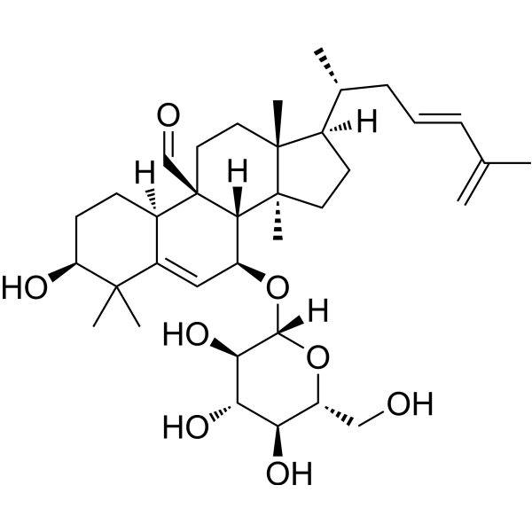
Kuguaglycoside C


| Product Name | Kuguaglycoside C |
| Price: | Inquiry |
| Catalog No.: | CN07672 |
| CAS No.: | 1041631-93-9 |
| Molecular Formula: | C36H56O8 |
| Molecular Weight: | 616.8 g/mol |
| Purity: | >=98% |
| Type of Compound: | Triterpenoids |
| Physical Desc.: | Powder |
| Source: | The fruits of Momordica charantia |
| Solvent: | DMSO, Pyridine, Methanol, Ethanol, etc. |
| SMILES: |
| Contact us | |
|---|---|
| First Name: | |
| Last Name: | |
| E-mail: | |
| Question: | |
| Description | Kuguaglycoside C is a triterpene glycoside that can be isolated from the leaves of Momordica charantia. Kuguaglycoside C induces caspase‐independent DNA cleavage and cell death of neuroblastoma cells. Kuguaglycoside C also significantly increases the expression and cleavage of apoptosis-inducing factor (AIF)[1]. |
| In Vitro | Kuguaglycoside C (0-100 μM, 48 h) 对 IMR-32 细胞具有显着的细胞毒性,IC50 为 12.6 μM[1]。 Kuguaglycoside C (30 μM, 48 h) 未显示 caspase-3 或 caspase-9 的激活,但显示 TRADD、AIF 和 DFF45 水平升高,以及 RIP1 和存活蛋白水平降低[1]. Hoechst 33342 染色检测显示高浓度 (100 μM) Kuguaglycoside C 诱导细胞核收缩,但流式细胞术未见凋亡小体[1]。 Cell Viability Assay[1] Cell Line: IMR-32 cells Concentration: 0, 0.3, 1, 3, 10, 30, 100 μM Incubation Time: 48 h Result: Induced significant cytotoxicity against the IMR-32 cells, with an IC50 of 12.6 μM. Western Blot Analysis[1] Cell Line: IMR-32 cells Concentration: 30 μM Incubation Time: 0, 2, 4, 8, 24, 48 h Result: Activated both type I (caspase-dependent) and type II (caspase-independent) DNases. Showed significant PARP cleavage. Showed increase in the levels of TRADD, AIF and DFF45, and decrease in the levels of RIP1 and survivin. Protein levels of cleaved caspase‐3, ‐7 and ‐9 were not increased. The expression levels of p‐p38 MAPK, p‐JNK, Bax, Bcl‐2 and cytochrome c remained unchanged. Cell Cycle Analysis[1] Cell Line: IMR-32 cells Concentration: 1, 3, 10, 30, 100 μM Incubation Time: 48 h Result: Had no effect on the cells of any phase of the cell cycle in the neuroblastoma cell line. |
| Density | 1.2±0.1 g/cm3 |
| Boiling Point | 734.0±60.0 °C at 760 mmHg |
| Flash Point | 222.4±26.4 °C |
| Exact Mass | 616.397522 |
| LogP | 5.18 |
| Vapour Pressure | 0.0±5.5 mmHg at 25°C |